
When most people think of skin cancer, they picture dark, irregular moles developing on sun-drenched shoulders, backs, or arms. Ultraviolet (UV) radiation from sun exposure is indeed the leading environmental risk factor for skin malignancies. However, the most aggressive form of skin cancer originates in pigment-producing cells called melanocytes, which exist throughout the human body.
Because melanocytes are present in places that rarely or never see sunlight, malignant tumors can form in hidden, non-exposed anatomical sites. These atypical lesions are often overlooked during routine self-examinations, leading to delayed diagnoses and more advanced stages at detection.
Understanding where skin cancer can hide, recognizing subtle warning signs, and conducting thorough head-to-toe skin checks are vital steps in early intervention.
Why Does Melanoma Develop in Hidden Areas?
While superficial spreading lesions typically develop on sun-exposed cutaneous surfaces, other subtypes arise independently of direct UV radiation. Genetic mutations, family history, cellular damage, and structural abnormalities within melanocytes can trigger malignant transformation anywhere pigment-producing cells reside.
The Problem with Hidden Lesions:
- Delayed Detection: Because these areas are out of sight, lesions often grow unnoticed until they produce secondary symptoms like pain, bleeding, or itching.
- Higher Risk of Metastasis: Late discovery increases the likelihood that cancer cells have invaded deeper tissue layers or regional lymph nodes.
- Diagnostic Complexity: Hidden lesions frequently mimic benign conditions such as fungal infections, bruises, blood blisters, or simple pigmentation changes.
7 Unexpected Places Melanoma Can Hide
A thorough self-examination requires looking beyond the usual areas. Dermatologists recommend checking these seven frequently overlooked anatomical sites:
[Head & Neck] ➔ Scalp, behind ears, mucosal tissues
[Extremities] ➔ Beneath nail beds (subungual), soles of feet, webbed spaces
[Hidden Folds] ➔ Underneath breasts, groin, perianal regions
1. The Scalp and Behind the Ears
Hair provides some natural shading, but it also obscures early-stage lesions. Scalp lesions are particularly dangerous because dense blood vessels and lymphatic networks in the head region allow cancer cells to spread rapidly if unchecked.
- What to look for: Raised bumps, dark spots, or patches that bleed easily when brushing hair.
2. Beneath the Nails (Subungual Melanoma)
Formally classified as acral lentiginous melanoma, subungual lesions develop under fingernails or toenails. They are more common in individuals with darker skin tones, who otherwise have lower rates of UV-induced skin cancers.
- What to look for: A narrow, dark brown or black vertical stripe running down the nail plate (Hutchinson’s sign), or persistent nail splitting that does not heal after injury.
3. Soles of the Feet and Between the Toes
The bottoms of the feet and the thin skin between toes receive minimal sunlight, yet they are prime locations for acral lesions.
- What to look for: Dark macules, unexplained sores that fail to heal, or spots that resemble stubborn plantar warts or bruises.
4. Mucosal Tissues (Inside the Mouth, Nose, and Passages)
Mucosal melanoma develops in the moist lining of internal passages, including the nasal cavities, oral mucosa, gastrointestinal tract, and genital regions. These represent less than 2% of all cases but carry a serious prognosis due to delayed identification.
- What to look for: Persistent sores inside the mouth, unexplained nosebleeds, or dark pigmented patches on mucosal membranes.
5. Skin Folds and Intimate Areas
Areas shielded from light such as the inframammary fold (under the breasts), the groin, and the perianal region are often excluded during quick self-checks.
- What to look for: New or changing moles, asymmetric spots, or persistent localized irritation.
6. The Eyes (Ocular Melanoma)
Melanocytes also exist in the uvea (the middle layer of the eye). Ocular lesions rarely show visible external symptoms in early stages and are usually discovered during routine dilated eye exams.
- What to look for: Blurred vision, sudden flashes of light, dark spots on the iris, or changes in pupil shape.
7. The Umbilicus (Belly Button)
The deep recesses of the belly button are easy to forget during routine skin audits, yet pigment changes can occur within these skin folds.
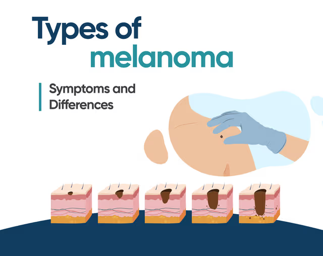
To better understand how these atypical presentations differ in appearance and cellular origin, explore the clinical overview of the different clinical types of melanoma, which details how nodular, acral, lentigo maligna, and superficial spreading variants behave.
How to Perform a Comprehensive Head-to-Toe Self-Exam?
Conducting a monthly self-examination helps establish a baseline for your skin, making it easier to spot new or changing spots.
| Body Zone | Examination Technique & Tools | Key Areas to Target |
| Head & Face | Blow-dryer to part hair; full mirror + hand mirror | Scalp, behind ears, inside ear cartilage, neck |
| Hands & Arms | Direct lighting; remove nail polish | Fingernails, nail beds, webbed skin between fingers |
| Torso & Folds | Full-length mirror; lift skin folds manually | Under breasts, belly button, sides of chest |
| Lower Body | Seated position; hand mirror for rear views | Soles of feet, toenails, heel edges, groin area |
The ABCDE Rules for Hidden Spots
While the standard ABCDE guide applies primarily to cutaneous surfaces, keeping these criteria in mind helps evaluate suspicious spots across all body regions:
- Asymmetry: One half of the spot does not match the other.
- Border: Edges are irregular, ragged, notched, or blurred.
- Color: Shade is non-uniform, combining shades of brown, black, pink, red, or blue.
- Diameter: Spot is larger than 6 millimeters (about the size of a pencil eraser), though some lesions present smaller.
- Evolving: Any mole or spot that changes in size, shape, color, or elevation over time, or begins to bleed, itch, or crust.
Frequently Asked Questions
Can you get melanoma in places that never see the sun?
Yes. While UV radiation causes the majority of cutaneous lesions, subungual, acral, and mucosal variants develop due to non-UV genetic factors, cellular errors, or inherited predisposition.
What does melanoma under a nail look like?
It usually presents as a dark brown or black line running vertically along the nail bed. Unlike a bruise caused by stubbing a toe or catching a finger, subungual dark bands do not grow out with the nail plate over time.
How often should I have a professional skin check?
Dermatologists generally recommend an annual professional skin examination for adults, or more frequently for individuals with a personal or family history of skin cancer, numerous dysplastic naevi, or prior significant sun exposure.
Summary
Early detection remains the single most effective factor in successfully treating skin cancer. While protecting sun-exposed skin with broad-spectrum sunscreen and protective clothing is essential, checking hidden areas such as the scalp, soles of the feet, nail beds, and skin folds is equally critical. By conducting regular full-body self-exams and scheduling annual professional skin checks, you can identify changing spots early, when treatment outcomes are most favorable.