
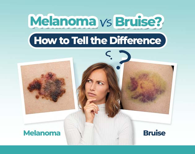
A dark spot or line under a toenail is a common concern. In most cases, subungual discoloration is simply a bruised toenail clinically known as a subungual hematoma caused by trauma or repeated friction. However, in rare instances, dark pigmentation beneath the nail plate can be a sign of subungual melanoma, a rare but serious form of skin cancer that originates in the nail matrix.
Because both conditions can present as brown, black, or purple discoloration, distinguishing between harmless bruising and a malignant lesion is critical for early detection and timely treatment.
What Is a Subungual Hematoma?
A subungual hematoma occurs when trauma causes small blood vessels under the nail bed to rupture, pooling blood beneath the hard nail plate.
Common Causes:
- Acute Trauma: Dropping a heavy object onto the toe or stubbing it forcefully.
- Repetitive Micro-Trauma: Running, hiking, or wearing tight-fitting footwear that creates constant pressure on the nail matrix.
Key Characteristics:
- Color: Ranges from reddish-purple to dark brown or black.
- Movement: As the nail grows outward over several months, the bruised area moves forward with it.
- Onset: Typically appears within 24 to 48 hours following an injury or strenuous physical activity.
- Sensation: Often accompanied by throbbing pain immediately after the injury, which diminishes over a few days.
What Is Subungual Melanoma?
Subungual melanoma is an acral lentiginous subtype of skin cancer that develops in the melanocytes (pigment-producing cells) within the nail matrix, the tissue beneath the cuticle that generates the nail plate. Unlike typical skin cancers, subungual melanoma is not primarily driven by sun exposure or ultraviolet (UV) radiation.
Key Characteristics:
- Appearance: Frequently presents as a longitudinal melanonychia a vertical brown, black, or dark stripe extending from the cuticle to the free edge of the nail.
- Progression: The pigment line remains stationary at the base or gradually widens, darkens, or changes shape over time without growing out with the nail plate.
- Hutchinson’s Sign: Pigmentation extending from the nail bed onto the adjacent skin of the cuticle or proximal nail fold is a classic diagnostic indicator of subungual malignancy.
- Sensation: Usually painless in its early stages, though advanced cases may cause nail dystrophy, splitting, or bleeding.
How to Monitor Toenail Discoloration at Home?
If you notice a dark mark beneath your toenail and are unsure of its origin, careful observation can help you gather helpful details before consulting a medical professional.
- Take Baseline Photographs: Capture a clear photo of the nail under good lighting alongside a small ruler or measurement reference.
- Track Growth Over 4 to 8 Weeks: Measure the distance between the cuticle and the bottom edge of the dark spot. If the clear gap between the cuticle and the spot increases over time, the discoloration is moving outward with nail growth, which strongly indicates a resolving bruise.
- Check Surrounding Skin: Inspect the cuticle and the fold of skin adjacent to the nail plate for any signs of hyperpigmentation.
- Note Physical Symptoms: Record whether the toe feels tender, throbbing, or completely pain-free.
Red Flag Symptoms That Require Medical Evaluation
While the vast majority of subungual dark spots are benign hematomas, specific signs warrant immediate clinical assessment by a dermatologist or podiatrist:
- A vertical dark band covering a single nail that broadens or darkens over time.
- Pigment spreads to the cuticle, side folds, or surrounding skin of the toe.
- Discoloration that remains in the exact same spot at the nail base for several months without moving forward.
- Unexplained splitting, cracking, or lifted areas of the nail plate without prior physical trauma.
- Spontaneous bleeding, ulceration, or discharge from beneath the nail.
When evaluating persistent or changing nail lesions, clinicians utilize specialized techniques such as dermoscopy, a non-invasive imaging method that examines micro-structures beneath the nail or a nail matrix biopsy to make a definitive diagnosis. Understanding these clinical diagnostic pathways and visual distinctions can provide valuable clarity; detailed breakdown guides on differentiating a melanoma or bruise on the toenail highlight the diagnostic markers clinicians evaluate during an examination.
Common Myths and Misconceptions
Myth 1:
“Subungual melanoma only affects fair-skinned individuals who get sunburns.”
Fact: Acral lentiginous melanoma occurs across all skin tones and is the most common subtype of melanoma diagnosed in individuals with darker skin tones. Because it develops in non-sun-exposed areas, UV exposure is not a primary risk factor.
Myth 2: “If there’s no pain, it’s nothing to worry about.”
Fact: Early-stage subungual melanoma is almost always pain-free. Pain is usually associated with acute trauma (bruising) or advanced malignant lesions that have invaded surrounding tissues.
Myth 3: “A bruised toenail should fade within a week.”
Fact: Toenails grow slowly typically about 1 to 2 millimeters per month. A complete toenail replacement can take anywhere from 9 to 12 months, meaning a deep hematoma may remain visible for up to a year as it slowly migrates toward the tip.
Frequently Asked Questions
How long does a bruised toenail take to grow out completely?
Toenails grow at an average rate of 1.5 to 2 millimeters per month. Depending on where the bruise is located, it can take anywhere from 6 to 12 months for a subungual hematoma to completely disappear as the nail plate grows outward.
Can a toenail bruise appear without an obvious injury?
Yes. Micro-trauma caused by tight footwear, downhill running, or repetitive pressure during sports can rupture small blood vessels under the nail without causing an immediate, memorable impact.
What does Hutchinson’s sign mean?
Hutchinson’s sign is the presence of brown or black pigment on the periungual skin (the cuticle or skin folds surrounding the nail). It is a classic clinical indicator of subungual melanoma and warrants prompt medical evaluation.
How do doctors confirm a subungual melanoma diagnosis?
A healthcare provider will first perform a visual examination using a dermatoscope. If the lesion exhibits suspicious features such as irregular pigmentation, varying band widths, or Hutchinson’s sign a biopsy of the nail matrix or nail bed is performed to analyze the tissue histologically.
Summary
Differentiating between a subungual hematoma and subungual melanoma comes down to tracking how the discoloration behaves over time. A bruised toenail usually follows a clear trauma, presents with initial pain, and gradually moves forward as the nail grows. In contrast, subungual melanoma typically manifests as a fixed dark vertical stripe, remains anchored at the nail matrix, and may involve surrounding skin. If you observe persistent discoloration that does not grow out, widens, or involves the cuticle, schedule an evaluation with a healthcare specialist for an accurate diagnosis.