
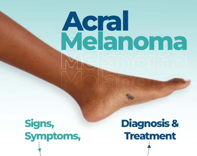
When most people think of skin cancer, they envision sun-damaged skin on the face, shoulders, or back. However, one of the most aggressive forms of skin cancer Acral Lentiginous Melanoma (ALM) develops on body parts that rarely see the sun. Famous reggae legend Bob Marley tragically passed away from this exact condition at age 36 after a dark spot under his toenail was initially mistaken for a soccer injury.
Because Acral Lentiginous melanoma forms in hidden, non-sun-exposed regions like the soles of the feet, the palms of the hands, and beneath fingernails or toenails, it is frequently misdiagnosed or caught in later stages.
In this comprehensive guide, you will learn what causes Acral Lentiginous melanoma, how to spot its early warning signs using the “CUBED” screening tool, how doctors diagnose and treat it, and what practical steps you can take to protect your health.
What is Acral Lentiginous Melanoma?
Acral Lentiginous Melanoma is a rare, distinct subtype of cutaneous melanoma that develops on the non-hair-bearing skin of the extremities.
To better understand the medical term:
- Acral: Derived from the Greek word akron, meaning highest or topmost point of the limbs (hands, feet, fingers, toes).
- Lentiginous: Refers to a freckle-like growth pattern where atypical pigment-producing cells (melanocytes) proliferate along the basal layer of the skin (the dermoepidermal junction).
- Melanoma: A severe form of cancer that originates in melanocytes.
Who Does It Affect?
While Acral Lentiginous melanoma accounts for only 2% to 5% of all new melanoma diagnoses overall, it represents the most common form of melanoma in individuals with darker skin phototypes. It makes up a disproportionate percentage of skin cancer cases among Black, Hispanic, and Asian populations. Because people with darker complexions often have a lower risk of sun-induced skin cancers, public awareness regarding acral screening remains dangerously low.
Recognizing the Symptoms: What Does ALM Look Like?
Because Acral Lentiginous melanoma does not follow typical sun-exposure patterns, recognizing its unique visual presentation is critical for early detection.
1. Palms and Soles
On the glabrous (hairless) skin of the hands and feet, early lesions typically appear as:
- An irregular, flat, or slightly raised dark patch or macule.
- Variegated coloring ranging from light tan and dark brown to jet black or blue-black.
- Asymmetrical borders that follow skin furrows and ridges.
- A growing spot that expands over months or years.
2. Subungual Melanoma (Under the Nails)
When Acral Lentiginous melanoma develops in the nail matrix, it presents as:
- A vertical, longitudinal dark band or streak running from the cuticle to the tip of the nail.
- A band that is wider than 3 millimeters or displays multiple shades of brown/black.
- Hutchinson’s Sign: A vital clinical clue where the dark pigment extends beyond the nail plate onto the surrounding skin fold or cuticle.
- Nail dystrophy, cracking, splitting, or lifting of the nail plate.
3. Amelanotic (Non-Pigmented) ALM
In roughly 10% to 20% of cases, Acral Lentiginous melanoma presents without melanin pigment. These lesions appear pink, red, or flesh-colored and are easily mistaken for benign conditions like plantar warts, non-healing foot ulcers, calluses, or fungal infections.
The “CUBED” Rule for Early Detection
The traditional “ABCDE” checklist used for general skin cancer screening (Asymmetry, Border, Color, Diameter, Evolving) often fails to detect acral lesions. Dermatologists instead recommend the specialized CUBED acronym for evaluating suspicious spots on the hands and feet:
- C – Colored: A lesion where any portion displays a color different from the surrounding normal skin.
- U – Uncertain Diagnosis: A spot or nail streak that lacks a clear, definitive clinical diagnosis.
- B – Bleeding: A lesion that bleeds, oozes fluid, or fails to scab over.
- E – Enlargement: A mole, patch, or band that is growing or deteriorating despite treatment.
- D – Delay: Any lesion that takes longer than two months to heal after injury or topical treatment.
Causes, Genetics, and Risk Factors
Unlike cutaneous melanoma, ultraviolet (UV) radiation from sunlight or tanning beds is not a primary cause of Acral Lentiginous melanoma.
Researchers are actively investigating the underlying mechanisms, but current evidence points toward a combination of physical and genetic drivers:
- Mechanical Stress & Shear Forces: The soles of the feet experience continuous weight-bearing pressure and friction, which may induce micro-trauma and stimulate abnormal cell growth.
- Distinct Molecular Driver Mutations: Genetic profiling shows that ALM has a lower overall tumor mutational burden than sun-induced melanomas. However, it frequently harbors specific gene amplifications and mutations, including KIT, NF1, CCND1 (Cyclin D1), TERT, and TP53.
- Age: Incidence increases sharply in adults aged 60 and older.
Diagnostic Procedures: How Doctors Evaluate ALM
Accurate, timely diagnosis requires specialized clinical tools and pathological confirmation.
Dermoscopy (Parallel Ridge Pattern)
A dermatologist uses a specialized handheld magnifier called a dermatoscope to examine the micro-patterns of skin pigment.
- Benign Acral Moles: Pigment aligns in the parallel furrows (grooves) of fingerprints or footprints.
- Acral Lentiginous Melanoma: Pigment concentrates preferentially along the parallel ridges of dermatoglyphics. Finding a Parallel Ridge Pattern on dermoscopy strongly indicates malignancy.
Biopsy and Histopathology
If a lesion is suspicious, a full-thickness biopsy is performed to confirm the diagnosis. Pathologists measure the Breslow depth (how deep the cancer has penetrated into the skin layer), check for cellular atypia, and evaluate whether ulceration is present.
Treatment Options for Acral Lentiginous Melanoma
Management depends on the stage at diagnosis, tumor thickness, and anatomical location.
1. Surgical Excision
Surgical removal remains the primary treatment for localized Acral Lentiginous melanoma:
- Wide Local Excision: The surgeon removes the primary tumor along with a safety margin of normal tissue (ranging from 0.5 cm for in-situ lesions to 2.0 cm for deeper invasive tumors). Reconstructive skin grafts or local flaps are often used to maintain hand or foot function.
- Digit-Sparing Nail Unit Excision: While historical treatment for subungual melanoma often required complete digit amputation, modern surgical protocols prioritize functional preservation through conservative, wide nail-unit excision whenever clinically safe.
2. Sentinel Lymph Node Biopsy (SLNB)
For tumors deeper than 0.8 mm to 1.0 mm or those with ulceration, doctors evaluate the nearest lymph node basin to check if cancer cells have begun to spread.
3. Systemic & Targeted Therapies
If the cancer has spread beyond its origin:
- Immunotherapy: Checkpoint inhibitor drugs (e.g., pembrolizumab, nivolumab) help the body’s immune system detect and attack cancer cells.
- Targeted Therapies: For tumors carrying KIT mutations, targeted kinase inhibitors (such as imatinib) can block cancer signaling pathways.
Frequently Asked Questions
Is Acral Lentiginous Melanoma caused by sun exposure?
No. Acral Lentiginous melanoma forms on areas that receive minimal UV light, such as the soles of the feet, palms, and under the nails. Sunscreen alone will not prevent this specific subtype.
What is the 5-year survival rate for Acral Lentiginous Melanoma?
When caught early in stage I or II, 5-year survival rates are high. However, the overall 5-year survival rate averages around 80% because many patients are diagnosed late after the lesion has already grown deep into the dermal tissue.
Can a dark line on my fingernail be a normal finding?
Yes. Dark longitudinal streaks (melanonychia) can occur naturally, especially in individuals with darker complexions. They can also result from trauma, fungal infections, or benign moles. However, any new, widening, or changing streak or one accompanied by skin discoloration around the cuticle should be evaluated immediately.
What is Hutchinson’s sign?
Hutchinson’s sign occurs when dark pigmentation from a subungual melanoma spreads from under the nail bed onto the surrounding skin, such as the cuticle or lateral nail fold. It is a key red-flag symptom requiring urgent evaluation.
Conclusion
Acral Lentiginous Melanoma proves that skin cancer checks should not stop at the neckline or shoulder blades. Because ALM can develop quietly beneath a fingernail or on the sole of your foot, routine self-examinations of your hands and feet are essential for catching changes early.